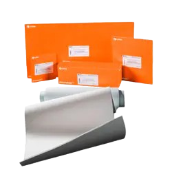
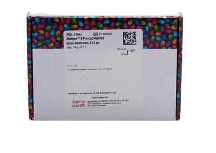
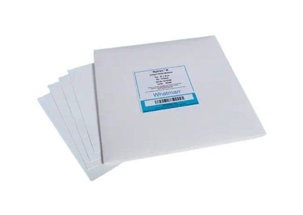
Western Blot and Nucleic Acid Membranes and Filter Papers
Suitable for Western blotting, dot-blot assays, and other protein or nucleic acid detection methods, including chemiluminescent, fluorescent, colorimetric, or radioactive techniques. Available in sheet, roll, or transfer stack formats, with various sizes and materials to suit your specific needs.

Useful Links
Save Now - Exclusive Deals

Product Code 18155995

Product Code 3604679

Product Code 4901753

Product Code 10168543

Product Code 200085296
Product Code 4880084

Product Code 3630927

Product Code 18104495
Must Have

Product Code 4696459

Product Code 4520455

Product Code 18791154

Product Code 18104984

Product Code 4525101


Complete Your Order - Great Deals

Product Code 10572913


Product Code 7226491
FAQ
SDS-PAGE and Western blot are two distinct but complementary techniques used in the analysis of proteins. The key differences are:
- SDS-PAGE separates proteins based on their molecular weight. Western blot detects specific proteins within a complex mixture after separation by SDS-PAGE
- SDS-PAGE uses sodium dodecyl sulfate (SDS) to denature proteins and impart a negative charge, allowing them to be separated by electrophoresis based on size. Western blot transfers proteins from an SDS-PAGE gel onto a membrane (such as PVDF or nitrocellulose), then uses specific antibodies to detect the protein of interest.
- SDS-PAGE sample preparation (including denaturation with SDS):
- Loading samples into the wells of a polyacrylamide gel
- Electrophoresis to separate proteins by size
- Staining the gel (e.g., with Coomassie Brilliant Blue or silver stain) to visualize all proteins
- Western blot sample preparation:
- Perform SDS-PAGE to separate proteins
- Transfer proteins from the gel to a membrane
- Block the membrane to prevent non-specific binding
- Incubate with a primary antibody specific to the target protein
- Incubate with a secondary antibody that binds to the primary antibody and is conjugated to a detectable enzyme or fluorophore
- Detect the target protein using appropriate detection methods (e.g., chemiluminescence, fluorescence)
- SDS-PAGE visualizes all proteins in the sample using a general protein stain. Western blot specifically detects the protein of interest using antibodies, providing information about the presence, size, and quantity of the target protein
- SDS-PAGE provides a profile of the protein mixture, showing the relative sizes and amounts of all proteins present. Western blot on the other hand provides specific information about the target protein, including its presence, molecular weight, and relative abundance
In summary, SDS-PAGE is primarily used for the separation of proteins by size, while Western blotting is used for the specific detection and analysis of individual proteins within a complex mixture after separation by SDS-PAGE.
The choice between PVDF (polyvinylidene difluoride) and nitrocellulose membranes in Western blotting depends on several factors:
Protein Binding Capacity:
PVDF has higher protein binding capacity, typically around 150-160 µg/cm². Nitrocellulose has lower protein binding capacity, typically around 80-100 µg/cm².
Durability:
PVDF is more durable and resistant to handling, making it suitable for multiple reprobing. Nitrocellulose is more fragile and prone to tearing, less suitable for multiple reprobing.
Sensitivity:
PVDF often provides higher sensitivity for detecting low-abundance proteins. Nitrocellulose is generally adequate for most applications, but might be less sensitive compared to PVDF.
Background Noise:
PVDF can sometimes exhibit higher background noise, which may require more stringent blocking and washing steps. Nitrocellulose typically shows lower background noise.
Compatibility with Staining:
PVDF is compatible with a variety of staining techniques, including Coomassie Brilliant Blue and Ponceau S. Nitrocellulose is also compatible with various staining techniques, but may not be as versatile as PVDF.
Cost:
PVDF is generally more expensive, while nitrocellulose is less expensive.
PVDF membranes are preferred for their higher protein binding capacity, durability, and sensitivity, especially when dealing with low-abundance proteins or requiring multiple reprobing. Nitrocellulose membranes are suitable for routine applications where cost is a concern and the proteins of interest are abundant, and they typically exhibit lower background noise. Choose based on the specific requirements of your experiment.
If a PVDF membrane dries out during the Western blotting process, several issues can arise:
- Loss of Protein Binding Capacity: Once the membrane dries, the proteins bound to it can become irreversibly attached in a way that may not be optimal for subsequent detection steps. This can lead to reduced sensitivity and inconsistent results
- Increased Background Noise: A dried PVDF membrane can exhibit higher background noise during detection, making it harder to distinguish the specific signal of the target protein
- Difficulty in Reprobing: If the membrane dries out, it can be more challenging to strip and reprobe it for other target proteins, as the stripping efficiency may be compromised
- Physical Damage: Drying out can make the membrane more brittle and prone to cracking or tearing, which can complicate handling and lead to loss of sample
To avoid these issues, it's crucial to keep the PVDF membrane moist throughout the entire Western blotting process. If the membrane does dry out, it can sometimes be rehydrated in methanol or water, but this may not fully restore its original functionality and performance.
Resources and Related Information


Shop By Brand